
Pericarditis

By the end of this post you will be able to:
- Recognize the clinical presentation of acute pericarditis and distinguish it from STEMI on ECG.
- Describe first-line pharmacological management, including colchicine, and the approach to recurrent pericarditis.
- Identify high-risk features that warrant admission and further workup.
TL;DR
What you need to know before seeing your first patient on your IM rotation with this condition
Estimated read time: 4 minutes

- What it is: Inflammation of the pericardium (the fibrous sac surrounding the heart). Most cases in developed countries are idiopathic or viral (>80% in outpatients). Other causes include autoimmune disease, post-cardiac injury syndrome (post-MI, post-cardiac surgery), malignancy (lung, breast, lymphoma), tuberculosis (common in endemic regions), and uremia.
- How it presents: Sharp, pleuritic chest pain that improves sitting up and leaning forward and worsens lying supine and with inspiration. Fever is common. A pericardial friction rub is reported in up to 1/3 of cases — a scratchy, high-pitched sound best heard with the diaphragm of the stethoscope at the lower left sternal border, with the patient leaning forward during held expiration. It may disappear once a pericardial effusion develops.
- Tests to order:
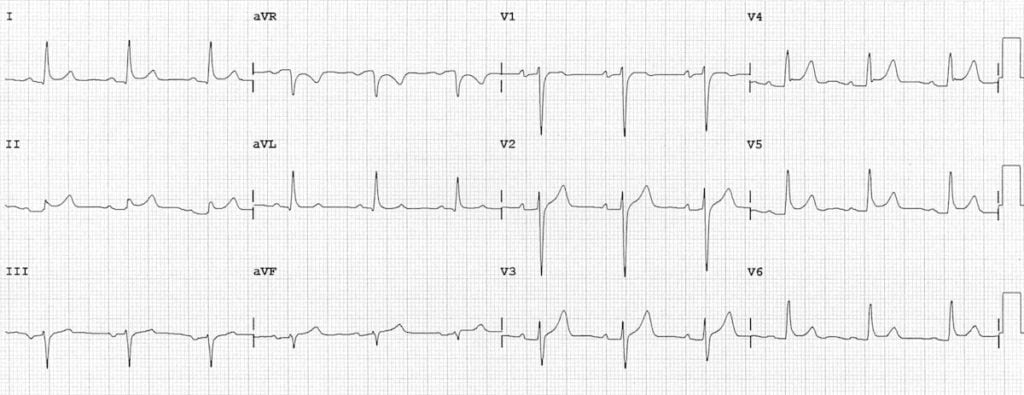
• ECG: Diffuse ST elevation + PR depression (see ECG below). Unlike STEMI, changes are diffuse (not territorial) and there are no reciprocal ST depressions
• Labs: CRP (elevated in 80–90%), CBC, troponin (mild elevation suggests myopericarditis), renal function, TSH
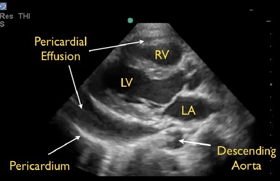
• Echo (TTE): Recommended for all patients (ESC 2025 Class I, first-line assessment). Assess for pericardial effusion (present in up to 60%, usually small), tamponade physiology, and rule out myocardial involvement
• Chest X-ray: Usually normal; enlarged cardiac silhouette if large effusion (>200 mL) - First-line treatment: NSAIDs + colchicine (dual therapy). Ibuprofen 600–800 mg TID or ASA 500–1000 mg TID, plus colchicine 0.5 mg BID (or 0.5 mg daily if <70 kg). Colchicine reduces recurrence by ~50%. GI upset (diarrhea, nausea) is the most common colchicine side effect — reduce to 0.5 mg daily if not tolerated. Add gastroprotection (PPI) with NSAIDs.
- Activity restriction: Limit exercise until symptoms resolve and CRP normalizes. Athletes should avoid competitive sport for at least 3 months (ESC 2025).
- When to admit: High-risk features include fever >38°C, large pericardial effusion, tamponade, failure to respond to NSAIDs within 7 days, elevated troponin (myopericarditis), immunosuppression, oral anticoagulant use, or trauma.
Pericarditis vs. STEMI on ECG
| Feature | Pericarditis | STEMI |
|---|---|---|
| ST elevation | Diffuse, concave (“smiley face”) | Territorial, convex (“frowny face”) |
| Reciprocal changes | None (except aVR, V1) | Present in opposite leads |
| PR segment | Diffuse PR depression | No PR changes |
| Q waves | Absent | May develop |
Listen: The Intern at Work

Read Around the Case
Based on the 2025 ESC Guidelines for Myocarditis and Pericarditis and the 2025 ACC Expert Consensus Statement on Pericarditis
Estimated read time: 12 minutes
Pathophysiology & Classification
The pericardium consists of two layers: a fibrous parietal layer and a serous visceral layer (epicardium) separated by 20–50 mL of serous fluid. Inflammation of these layers produces the clinical syndrome of pericarditis — chest pain from friction between inflamed surfaces, effusion from increased fluid production, and ECG changes from inflammation extending to the subepicardium.
The ESC 2025 guidelines classify pericarditis by duration:
- Acute pericarditis: ≤4 weeks duration. Most common presentation.
- Incessant pericarditis: Symptoms persist >4 weeks but <3 months without remission.
- Recurrent pericarditis: Documented recurrence after a symptom-free interval of ≥4 weeks. Occurs in 15–30% of patients after a first episode; risk increases without colchicine and with corticosteroid use.
- Chronic pericarditis: >3 months duration.
Etiologies
Diagnostic Criteria (ESC 2025 / ACC 2025)
The diagnosis of acute pericarditis requires a clinical presentation (pleuritic chest pain or equivalent) plus ≥1 additional finding:
- Pericardial friction rub on auscultation (≤33% of cases)
- ECG changes: diffuse ST elevation, PR depression
- Elevated inflammatory markers: CRP (≥80% of cases), ESR
- Pericardial effusion new or worsening on imaging (echo preferred)
- Pericardial inflammation on CMR (late gadolinium enhancement) or CT
The ACC 2025 consensus grades certainty: 0 additional findings = unlikely; 1 = possible; ≥2 = definite diagnosis.
ECG Evolution in Pericarditis
ECG changes in pericarditis reflect subepicardial inflammation and evolve through four classic stages, though not all patients demonstrate every stage:
The ESC 2025 notes that ECG changes imply concomitant subepicardial inflammation and are more common in younger patients. They are reported in 20–60% of cases. Atrial fibrillation can also occur, especially in predisposed patients.
Risk Stratification & Triage
Most patients with idiopathic/viral acute pericarditis can be managed as outpatients. The ESC 2025 identifies the following high-risk features (red flags) that should prompt admission and further workup:
- Fever >38°C
- Large pericardial effusion (>20 mm on echo) or tamponade
- Elevated troponin (suggests myopericarditis)
- Failure to improve after ≥7 days of NSAID therapy
- Immunosuppression
- Oral anticoagulant use (risk of hemorrhagic effusion)
- Acute trauma
- Subacute onset without clear inflammatory features (consider malignancy, TB)
Pharmacological Management
The cornerstone of treatment is empirical dual anti-inflammatory therapy: an NSAID (or aspirin) at full anti-inflammatory doses plus colchicine. The ESC 2025 recommends gastroprotection with a PPI when using NSAIDs.
Taper NSAIDs only after symptom resolution AND CRP normalization. Colchicine does not require tapering (ACC 2025 notes tapering “may be considered”). Monitor CRP to guide treatment length.
Colchicine: Side Effects & Interactions
GI side effects are the most common reason for discontinuation: diarrhea (~10%), nausea, vomiting, abdominal cramping. Taking colchicine with food may reduce GI intolerance. Dose reduction to 0.5 mg daily often resolves symptoms.
- Drug interactions: Colchicine is metabolized by CYP3A4 and excreted via P-glycoprotein. Avoid concurrent use with strong CYP3A4 inhibitors (clarithromycin, ketoconazole, HIV protease inhibitors) — risk of severe toxicity including bone marrow suppression. Reduce dose with moderate inhibitors (diltiazem, verapamil).
- Renal impairment: Use 0.5 mg daily if eGFR <30 mL/min. Contraindicated in dialysis patients receiving strong CYP3A4 inhibitors.
- Rare toxicities: Bone marrow suppression (pancytopenia), myopathy, neuropathy — usually with overdose or drug interactions. Monitor CBC if prolonged use.
Recurrent Pericarditis
Recurrence occurs in 15–30% of patients and is the most challenging aspect of pericarditis management. Risk factors include incomplete treatment of the first episode, early corticosteroid use, and not using colchicine.
The management ladder (ESC 2025 / ACC 2025):
- First-line: NSAIDs/ASA + colchicine (same as acute episode, but colchicine for ≥6 months)
- Second-line: Low-dose corticosteroids (0.2–0.5 mg/kg/day prednisone) + colchicine, or triple therapy (NSAID + corticosteroid + colchicine) for refractory cases
- Third-line (corticosteroid-dependent): Anti-IL-1 agents — anakinra (100 mg SC daily) or rilonacept (loading 320 mg SC, then 160 mg SC weekly). The AIRTRIP trial demonstrated anakinra significantly reduced recurrence in colchicine-resistant, corticosteroid-dependent patients.
- Last resort: Pericardiectomy for medically refractory cases at an expert surgical centre
Complications
Pericardial effusion is present in up to 60% of acute pericarditis cases, usually small and hemodynamically insignificant. Effusions are classified by size on echo: small (<10 mm), moderate (10–20 mm), large (>20 mm).
- Constrictive pericarditis: Rare complication (<1% of idiopathic/viral pericarditis, higher with TB or purulent). Fibrotic thickening of pericardium impairs diastolic filling → right heart failure (edema, ascites, elevated JVP with Kussmaul sign). Diagnosis via echo, CT, CMR, and cardiac catheterization. Treatment is pericardiectomy for advanced cases; anti-inflammatory therapy for transient constriction.
- Myopericarditis: Troponin elevation with pericarditis indicates concomitant myocardial involvement. Generally preserves LV function and has a benign prognosis if LVEF is normal. Avoid NSAIDs if significant LV dysfunction (use colchicine alone).
Cardiac tamponade occurs when pericardial fluid accumulation compresses the cardiac chambers, impairing diastolic filling and reducing cardiac output. The rate of accumulation matters more than the absolute volume — rapid accumulation of even 150–200 mL can cause tamponade, while slow accumulation may tolerate >1 L. Requires urgent echo-guided pericardiocentesis.
sounds
neck veins
Note: The full triad is present in a minority of cases. Tachycardia and tachypnea are often the earliest signs. Electrical alternans on ECG (beat-to-beat QRS amplitude variation) is highly specific but insensitive.
Definition: A >10 mmHg drop in systolic blood pressure during normal inspiration. Measured with a manual sphygmomanometer: deflate slowly, note the pressure at which Korotkoff sounds are first heard only during expiration, then continue deflating until sounds are heard throughout the respiratory cycle — the difference between these two pressures is the pulsus paradoxus.
Mechanism: During inspiration, negative intrathoracic pressure increases venous return to the right heart. In tamponade, the fixed pericardial volume means increased RV filling causes the interventricular septum to bow into the LV, reducing LV filling and stroke volume — producing an exaggerated drop in systolic BP.
Clinical tip: Pulsus paradoxus may be absent in tamponade with severe aortic regurgitation, atrial septal defect, or severe LV dysfunction. It can also be present in severe asthma, COPD, and tension pneumothorax.
Avoid corticosteroids as first-line therapy for idiopathic/viral pericarditis. Multiple trials (COPE, CORP-2) have shown that corticosteroid use — especially at high doses or with rapid tapers — increases the risk of recurrence. Reserve corticosteroids for autoimmune etiologies, true NSAID contraindications, or refractory cases. When corticosteroids are necessary, use the lowest effective dose (prednisone 0.2–0.5 mg/kg/day) and taper very slowly.
Test Yourself
Click on your answer to reveal the explanation.
A 28-year-old previously healthy man presents to the emergency department with 2 days of sharp chest pain that worsens when lying flat and improves when sitting forward. His temperature is 37.8°C, HR 88, BP 126/78. ECG shows diffuse concave ST elevation with PR depression in multiple leads. CRP is elevated at 85 mg/L. Troponin is normal. Echo shows a small pericardial effusion.
What is the most appropriate initial treatment?
A 45-year-old woman presents with her third episode of pericarditis in 18 months. She initially responded to ibuprofen and colchicine but symptoms recurred each time colchicine was stopped at 3 months. She is currently on prednisone 15 mg daily (started 6 weeks ago for the current flare) and colchicine 0.5 mg BID, with improving symptoms and a CRP trending down from 60 to 12 mg/L.
After reversible triggers have been addressed, what is the most appropriate management?
Reflect
A patient with acute pericarditis asks you why you’re prescribing colchicine — a “gout medication” — for a heart condition. How would you explain, in plain language, what colchicine does for pericarditis and why it’s important to take it for the full 3 months even after feeling better?
Please do not include patient identifying details — no names, initials, dates, medical record numbers, or any detail that could identify a specific patient or encounter. Write about your reasoning, not about a real person.
Further Reading
-
Guideline
2025 ESC Guidelines for the Management of Myocarditis and Pericarditis — Schulz-Menger & Imazio et al. Eur Heart J 2025. -
Expert Consensus
2025 ACC Expert Consensus Statement on the Diagnosis and Management of Pericarditis — Wang & Klein et al. JACC 2025. -
Landmark Trial
Colchicine as First-Choice Therapy for Recurrent Pericarditis (CORP-2) — Imazio et al. Ann Intern Med 2014;160:556–564. -
Landmark Trial
Colchicine for Acute Pericarditis (ICAP) — Imazio et al. NEJM 2013;369:1522–1528. -
Landmark Trial
Anakinra for Corticosteroid-Dependent Recurrent Pericarditis (AIRTRIP) — Brucato et al. Lancet 2016;387:1855–1862.