
Infective Endocarditis

By the end of this post you will be able to:
- Apply the Modified Duke Criteria, including the correct approach to blood cultures and when a negative TTE warrants a TEE.
- Select empiric antibiotic therapy for suspected infective endocarditis.
- Identify the indications for surgical management.
TL;DR
What you need to know before seeing your first patient on your IM rotation with this condition
Estimated read time: 3 minutes

- What it is: Infection of the heart valves or endocardial surface. Most commonly caused by Staphylococcus aureus (now the #1 cause in developed countries), streptococci, or enterococci.
- How it presents: Fever + new or changing murmur is the classic combination. Also look for: embolic phenomena (stroke, splenic infarcts, Janeway lesions), immunologic phenomena (Osler nodes, Roth spots, glomerulonephritis), and persistently positive blood cultures.
- Blood cultures first — always: Obtain 3 sets from separate venipuncture sites before antibiotics. IE bacteremia is continuous — do NOT wait for fever spikes.
- Initial workup:
• ECG: New PR prolongation or bundle branch block → suspect perivalvular abscess
• Labs: CBC, CRP/ESR, creatinine, urinalysis (hematuria from immune complex GN)
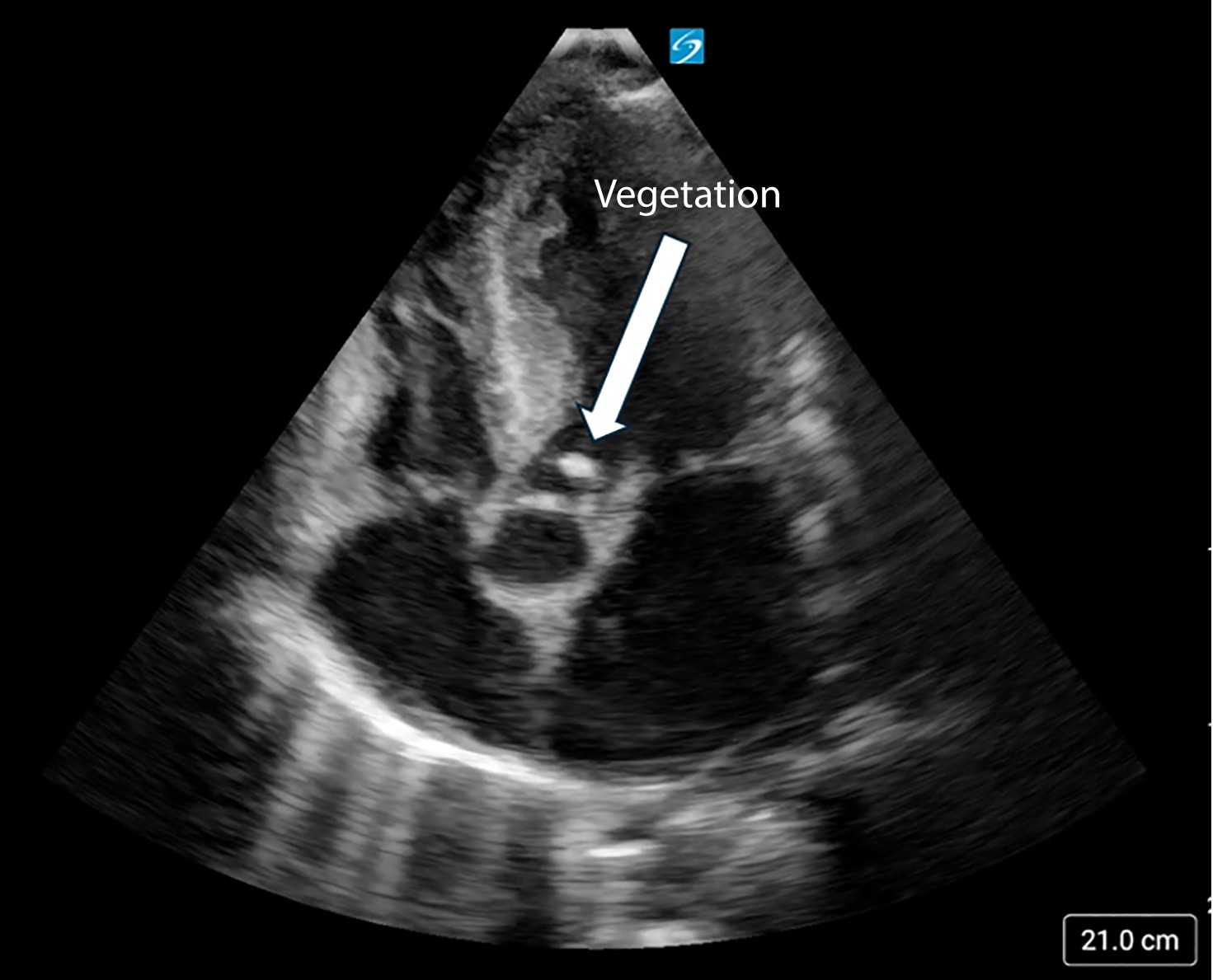
• Echo: TTE is first-line, but sensitivity is only ~70% (native valve) and ~50% (prosthetic). A negative TTE does not rule out IE — if clinical suspicion remains, escalate to TEE - Modified Duke Criteria: Definite IE = 2 major criteria, or 1 major + 3 minor, or 5 minor. Major criteria: (1) typical organisms on blood culture, (2) endocardial involvement on echo.
- Empiric therapy: Vancomycin + ceftriaxone (native valve); add gentamicin + rifampin for prosthetic valves.
- When to call for help: Hemodynamic instability, embolic events, suspected abscess, prosthetic valve IE, or S. aureus bacteremia not clearing within 72 hours.
Listen: The Intern at Work

Read Around the Case
Based on the 2015 AHA Scientific Statement and the 2023 ESC Guidelines for IE Management
Estimated read time: 14 minutes
Pathophysiology & Classification
IE begins with endothelial damage (from turbulent flow, devices, or prior valvular disease), leading to deposition of fibrin and platelets — a nonbacterial thrombotic endocarditis (NBTE). During bacteremia, organisms adhere to this nidus and proliferate within a biofilm-like vegetation, protected from host defenses. The vegetation can cause local valve destruction, embolic events, and systemic immune complex disease.
- Native valve IE (NVE): Most common. Mitral > aortic. S. aureus is now the leading cause in developed countries.
- Prosthetic valve IE (PVE): Early (<60 days post-op) — typically nosocomial organisms (CoNS, S. aureus, gram-negatives). Late (>60 days) — similar organisms to NVE.
- CIED-related IE: Pacemakers, ICDs. Lead extraction usually required. Prolonged antibiotics.
- Right-sided IE: Predominantly tricuspid valve, seen in IVDU. S. aureus is the most common cause. Lower mortality than left-sided IE.
Modified Duke Criteria
The modified Duke criteria remain the standard diagnostic framework. Definite IE = 2 major, or 1 major + 3 minor, or 5 minor. Possible IE = 1 major + 1 minor, or 3 minor.
Blood Cultures & Diagnostic Workup
3 sets of blood cultures from separate venipuncture sites before antibiotics — this is the single most important diagnostic step (AHA Class I, Level A). The first and last sets should be drawn at least 1 hour apart. IE bacteremia is continuous; do NOT wait for fever spikes.
Culture-negative IE (5–10% of cases) — consider: prior antibiotics (most common cause), HACEK organisms (slow-growing), Coxiella burnetii (Q fever), Bartonella species, Brucella, Tropheryma whipplei, fungi. Request serologies and PCR when standard cultures are negative.
- Labs: CBC, CRP/ESR, renal function, urinalysis (hematuria from immune complex GN)
- ECG: New conduction abnormality (PR prolongation, new bundle branch block) raises concern for perivalvular abscess
- Echo: See TTE vs TEE section below
Imaging: TTE vs TEE
Transthoracic echocardiography (TTE) is the first-line imaging study — it is non-invasive and can be done at the bedside. However, TTE sensitivity is only ~70% for native valves and ~50% for prosthetic valves (AHA 2015). Transesophageal echocardiography (TEE) provides superior image resolution because the probe is positioned in the esophagus, directly behind the heart. TEE is significantly more sensitive for vegetations, abscesses, and prosthetic valve complications, but it requires conscious sedation and carries a small risk of esophageal injury.
Get TEE when:
- TTE is negative but clinical suspicion remains (especially with S. aureus bacteremia)
- Prosthetic valve or CIED
- Suspected complications (abscess, fistula, dehiscence)
- Poor TTE windows (obesity, COPD, post-surgical)
Repeat imaging in 5–7 days if initial TTE/TEE is negative but suspicion persists. The 2023 ESC guidelines also recommend cardiac CT and 18F-FDG PET/CT as complementary imaging modalities, particularly for prosthetic valve and CIED-related IE.
Step 1: TTE for all suspected IE
Step 2: If TTE is negative but clinical suspicion remains → TEE
Step 3: If both negative → repeat in 5–7 days
Go straight to TEE if: prosthetic valve, CIED, S. aureus bacteremia, or suspected abscess
Empiric Antibiotic Therapy
Start empiric therapy after blood cultures are drawn. Regimen selection depends on whether the valve is native or prosthetic, and local resistance patterns. Infectious diseases consultation should occur at initiation of empiric therapy (AHA 2015).
Narrow therapy once organism and susceptibilities are identified. Vancomycin troughs: 15–20 μg/mL (or AUC/MIC-guided dosing).
Definitive Antibiotic Therapy by Organism
PVE generally requires longer therapy (6 weeks minimum). Add rifampin for staphylococcal PVE. Gentamicin: monitor levels (peak 3–4 μg/mL, trough <1 μg/mL) and renal function. Ampicillin + ceftriaxone preferred over ampicillin + gentamicin for enterococcal NVE to avoid nephrotoxicity.
IV-to-Oral Antibiotic Switch
The POET trial (NEJM 2019) demonstrated that switching to oral antibiotics after ≥10 days of IV therapy was non-inferior to completing the full IV course in patients with left-sided IE. This has been incorporated into the 2023 ESC guidelines.
Eligibility criteria for oral switch:
- ≥10 days of IV antibiotics completed
- Afebrile for ≥48 hours
- Blood cultures negative
- No abscess or perivalvular extension
- No signs of heart failure
- No indication for surgery
- Applicable organisms: streptococci, E. faecalis, S. aureus, CoNS
Oral regimens should use high-dose agents with good bioavailability (e.g., amoxicillin 1 g TID, linezolid, moxifloxacin, rifampin), guided by susceptibilities. The total antibiotic duration remains the same (4–6 weeks).
Early surgical consultation improves outcomes. The AHA identifies the following indications:
- Heart failure from valvular dysfunction (most common indication for surgery)
- Uncontrolled infection: persistent bacteremia >5–7 days on appropriate therapy, enlarging vegetation, abscess, fistula, or pseudoaneurysm
- Prevention of embolism: vegetation >10 mm with embolic event, or >15 mm isolated (especially anterior mitral leaflet)
- Prosthetic valve IE caused by S. aureus or fungi — high mortality with medical therapy alone
- New conduction abnormality suggesting perivalvular abscess extension
The EASE trial (Kang et al., NEJM 2012) showed that early surgery within 48 hours reduced the composite endpoint of death and embolic events from 28% to 3% at 6 months.
The ESC 2023 guidelines use a hub-and-spoke model: uncomplicated IE can stay local with early, regular communication with a reference centre Endocarditis Team (Class I, Level B). Complicated IE should be transferred early to a centre with surgical facilities (Class IIa, Level B).
In Canada, phone-based specialist consultation is available through programmes like RACE (BC) and Specialist LINK (Alberta), which connect community physicians with cardiology, ID, and cardiac surgery in real time.
Complications
- Embolic events (20–50%): stroke, splenic/renal infarcts, mycotic aneurysms. Highest risk in first 2 weeks of therapy; declines significantly after initiation of effective antibiotics.
- Perivalvular abscess: Suspect when fever persists despite appropriate antibiotics + new conduction abnormality on ECG. More common with aortic valve IE. TEE is the imaging modality of choice.
- Heart failure: Most common cause of death. Usually from acute valvular regurgitation. Urgent surgical consultation.
- Mycotic aneurysms: Consider CT angiography if neurologic symptoms develop. Most intracranial mycotic aneurysms are in the distal branches of the MCA.
Special Populations
- IVDU: Right-sided (tricuspid) IE predominates. S. aureus is the most common pathogen. Lower threshold for empiric coverage. The CJC 2025 Canadian Working Group recommendations address practical management considerations in this population.
- Prosthetic valve: Higher mortality (>20%). Broader empiric coverage. Longer treatment (≥6 weeks). Lower threshold for TEE and surgery. Add rifampin for staphylococcal PVE.
- CIED-related: Lead extraction is usually required in addition to prolonged antibiotics (AHA Class I). Reimplantation at a new site after blood cultures are negative.
- S. aureus bacteremia: All SAB patients need echocardiography. The CMAJ 2019 SAB review recommends a minimum of 14 days IV therapy for uncomplicated SAB and 4–6 weeks for complicated SAB or IE.
Antibiotic prophylaxis before dental procedures is recommended only for patients at highest risk of IE (AHA 2015, Class IIa). This includes:
- Prosthetic heart valves (mechanical or bioprosthetic)
- Previous IE
- Certain congenital heart disease: unrepaired cyanotic CHD, repaired with prosthetic material within 6 months of procedure, or repaired with residual defects adjacent to prosthetic material
- Cardiac transplant recipients with valvulopathy
Procedures requiring prophylaxis: dental procedures involving manipulation of gingival tissue, the periapical region of teeth, or perforation of the oral mucosa. Prophylaxis is not recommended for GI or GU procedures.
Regimen: Amoxicillin 2 g PO, 30–60 minutes before the procedure. If unable to take oral medication: ampicillin 2 g IV/IM. Penicillin allergy: clindamycin 600 mg PO (or IV).
Every patient with S. aureus bacteremia needs an echo. SAB carries a 25–30% risk of IE. A negative TTE does not rule it out — TEE should be performed if there is persistent bacteremia (>72h), a prosthetic valve, CIED, or ongoing clinical concern. Never assume SAB is “just a line infection” without adequate evaluation.
Related Trial Files
Early Surgery vs. Conventional Treatment for IE (EASE Trial)
Trial Files Throwback →

Listen: Deep Dives
Test Yourself
Click on your answer to reveal the explanation.
A 42-year-old man with a history of injection drug use presents with five days of fever, night sweats and malaise. He has no localizing symptoms and no prior cardiac history. Temperature is 38.9 °C, heart rate 105 beats/min, blood pressure 118/72 mmHg, respiratory rate 18, oxygen saturation 96% on room air. A new holosystolic murmur is audible at the left lower sternal border, louder on inspiration. There are no peripheral stigmata. He is alert, well perfused and not in shock.
Which of the following is the most appropriate next step?
A 58-year-old man with native mitral valve endocarditis due to Streptococcus gallolyticus has completed ten days of intravenous ceftriaxone. He was never hypotensive and has had no embolic events. Blood cultures have been negative for seven days and he has been afebrile for five. Repeat transesophageal echocardiography shows a stable 8 mm vegetation with no abscess or perivalvular extension. He has no signs of heart failure and no indication for surgery. Renal function is normal and he has reliable oral intake.
Which of the following is the most appropriate next step?
Reflect
Your team gets a normal TTE back on a patient with Staph aureus bacteremia. The senior resident says, “TTE is negative — we can rule out endocarditis.” Do you agree? Why or why not?
Please do not include patient identifying details — no names, initials, dates, medical record numbers, or any detail that could identify a specific patient or encounter. Write about your reasoning, not about a real person.
Further Reading
-
Guideline
2015 AHA Scientific Statement: Infective Endocarditis in Adults — Baddour et al. Circulation 2015;132:1435–1486. -
Guideline
2023 ESC Guidelines for the Management of Endocarditis — Delgado et al. Eur Heart J 2023;44:3948–4042. -
Canadian Resource
The Evolving Landscape of Infective Endocarditis: Past, Present, and Future — CJC 2025. -
CMAJ
The Risk of Infective Endocarditis Among People Who Inject Drugs — CMAJ 2019;191:E93. -
Landmark Trial
Partial Oral versus IV Antibiotic Treatment of Endocarditis (POET Trial) — Iversen et al. NEJM 2019;380:415–424.