
Coronary Artery Disease

By the end of this post you will be able to:
- Recognize the spectrum of acute coronary syndromes and differentiate STEMI from NSTEMI on ECG.
- Initiate acute management of STEMI (reperfusion strategy) and NSTEMI (risk stratification and timing of invasive strategy).
- Prescribe post-ACS secondary prevention, including DAPT and guideline-directed medical therapy.
- Diagnose and manage stable angina with anti-anginal therapy and appropriate investigations.
TL;DR
What you need to know before seeing your first patient on your IM rotation with this condition
Estimated read time: 5 minutes


- The ACS spectrum: Coronary artery disease (CAD) presents acutely as acute coronary syndrome (ACS) — a spectrum from unstable angina to NSTEMI to STEMI. The key distinction is whether there is ST elevation on ECG (STEMI) or not (NSTEMI/unstable angina), and whether troponin is elevated (MI vs unstable angina). Stable angina is predictable chest pain with exertion that resolves with rest — not an emergency.
- Does this patient with chest pain have ACS? (JAMA Rational Clinical Exam, Fanaroff 2015). Likelihood ratios (LR) above 1 argue for ACS, below 1 against:
• ST depression on ECG (LR 5.3) — the single strongest predictor
• Pain radiating to the right arm (LR 2.9) or both arms (LR 2.6)
• History of peripheral arterial disease (LR 2.7) or a prior abnormal stress test (LR 3.1)
• Associated diaphoresis (LR 2.0); pain described as “pressure” (LR 1.3 — classic but weak)
• Pain reproducible by palpation (LR 0.3) makes ACS less likely but does not exclude it
• No single finding rules out ACS — ~13% of ED chest pain patients ultimately have one - ECG patterns — STEMI vs NSTEMI:
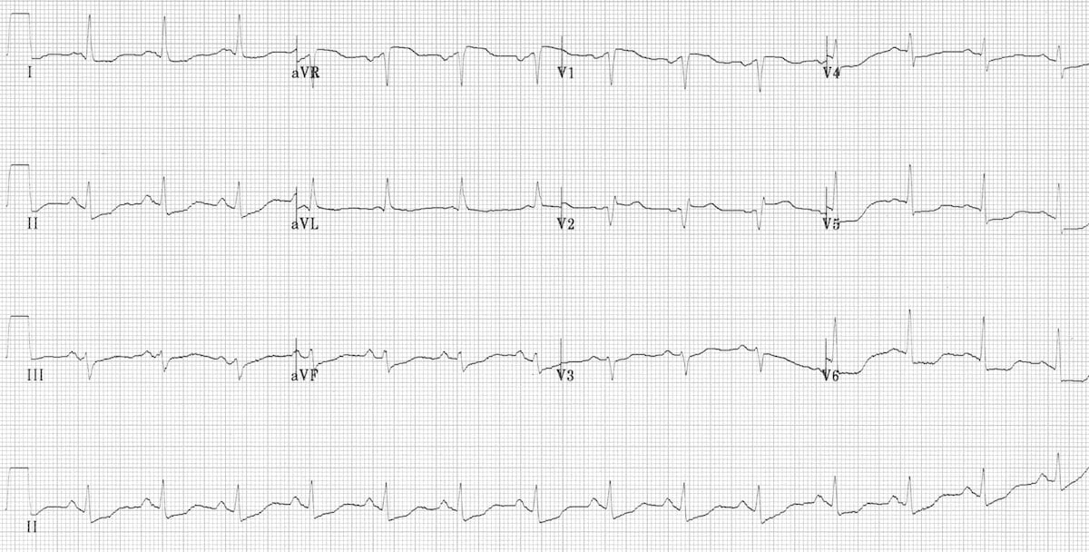
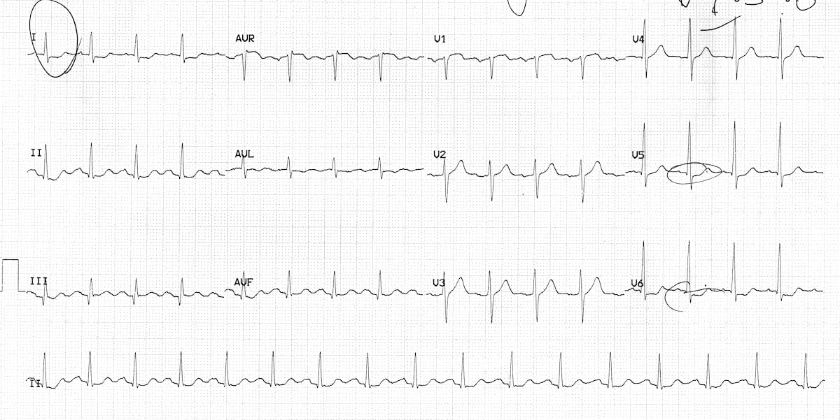
ECG images from LITFL ECG Library, used under CC BY-NC-SA 4.0.
- Initial investigations: Order immediately when you suspect ACS:
• Stat 12-lead ECG (within 10 minutes of presentation) — repeat in 15–30 min if initial is non-diagnostic
• Troponin (high-sensitivity cTnT or cTnI) — at presentation and repeat at 3–6 hours. A rising pattern (“delta”) confirms acute myocardial injury
• CBC (anemia worsens ischemia), electrolytes + creatinine (renal function for contrast/drugs), glucose, coagulation studies (INR/PTT before anticoagulation)
• CXR (rule out pulmonary edema, aortic dissection), lipid panel - Initial management of STEMI — Time = Muscle:
• ASA 160–325 mg chewed immediately
• Activate the cath lab for primary PCI — target door-to-balloon <90 minutes (CCS 2019)
• If PCI not available within 120 min of first medical contact: fibrinolysis (door-to-needle <30 min), then transfer for angiography within 24 hours
• P2Y12 inhibitor (ticagrelor or clopidogrel) given in the ED or cath lab
• Heparin (UFH) for procedural anticoagulation - Initial management of NSTEMI:
• ASA 160–325 mg chewed immediately
• Risk stratify — high-risk features include ongoing chest pain, hemodynamic instability, new HF, dynamic ECG changes, or rising troponin
• High-risk patients: early invasive strategy (angiography within 24 hours)
• Lower-risk patients: can be managed conservatively initially with medical therapy, with angiography within 24–72 hours
• P2Y12 inhibitor: CCS 2023 recommends not pretreating NSTEMI patients with P2Y12 ≤24h before angiography (give at time of PCI)
• Anticoagulation (UFH or enoxaparin) until angiography
Listen: MedicinePods

Read Around the Case
Based on CCS 2019 STEMI Guidelines, CCS 2023 Antiplatelet Therapy Update, and CCS 2014 Stable Ischemic Heart Disease Guidelines
Estimated read time: 19 minutes
1. Presentation & Diagnosis
ACS represents a continuum of acute myocardial ischemia caused by plaque rupture or erosion with superimposed thrombus formation. The initial ECG divides the pathway:
ACS Classification
Stat ECG within 10 minutes
→ STEMI
Emergent reperfusion
Troponin positive → NSTEMI
Troponin negative → Unstable Angina
ECG Interpretation in ACS
- STEMI criteria: New ST elevation at the J-point in ≥2 contiguous leads: ≥2 mm (men ≥40 years) or ≥2.5 mm (men <40) in V2–V3; ≥1.5 mm (women) in V2–V3; ≥1 mm in all other leads
- STEMI equivalents (don’t miss these):
• De Winter T-waves: Upsloping ST depression with tall, symmetric T-waves in precordial leads (LAD occlusion without ST elevation)
• Wellens syndrome: Deeply inverted or biphasic T-waves in V2–V3 during pain-free interval (critical proximal LAD stenosis)
• Posterior MI: ST depression in V1–V3 (mirror image); confirm with posterior leads V7–V9
• New LBBB with clinical suspicion — apply Sgarbossa criteria - NSTEMI patterns: ST depression (≥0.5 mm), T-wave inversion, or even a normal ECG. A normal ECG does not exclude ACS — serial ECGs and troponin are essential.
- Reciprocal changes increase specificity for STEMI (e.g., ST depression in aVL with inferior STEMI)
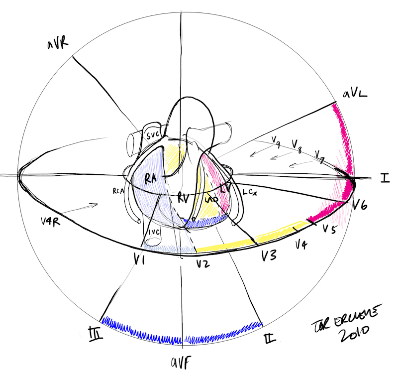
Diagram by Dr Tor Ercleve, from the LITFL MI Localization library, used under CC BY-NC-SA 4.0.
High-Sensitivity Troponin Interpretation
- hs-cTnT <5 ng/L at presentation: Very high negative predictive value for MI (>99%). Consider 1-hour rule-out algorithm (0h/1h) where available.
- Rising pattern (delta): A rise or fall of ≥20% with at least one value above the 99th percentile confirms acute myocardial injury (MI). A static elevation suggests chronic myocardial injury (e.g., CKD, HF).
- Troponin is not specific for ACS: Elevated troponin = myocardial injury, not necessarily coronary thrombosis. Consider: PE, myocarditis, takotsubo, sepsis, type 2 MI (demand ischemia), renal failure.
Type 1 vs Type 2 MI: Type 1 MI is caused by atherothrombotic plaque rupture/erosion — this is “classic” ACS requiring antiplatelet therapy and consideration of angiography. Type 2 MI is caused by supply-demand mismatch (tachycardia, anemia, hypotension, hypoxia) without plaque rupture — treat the underlying cause, not the troponin. This distinction changes management entirely.
2. STEMI Management
The CCS 2019 STEMI guideline emphasizes rapid reperfusion within 12 hours of symptom onset as the cornerstone of STEMI care. Canada uses a hub-and-spoke model with regional STEMI networks.
STEMI Reperfusion Strategy (CCS 2019)
ECG within 10 min of first medical contact (FMC)
Primary PCI
Door-to-balloon <90 min (at PCI centre)
FMC-to-device <120 min (with transfer)
Fibrinolysis
Door-to-needle <30 min
Then transfer for angiography within 24h
Adjunctive Therapy in STEMI (CCS 2019)
- ASA 160–325 mg chewed at FMC (Strong Recommendation)
- P2Y12 inhibitor: ticagrelor 180 mg or clopidogrel 600 mg load — give in the ED or cath lab, not prehospital
- Anticoagulation: UFH for the procedure (Strong); bivalirudin if HIT or very high bleeding risk
- Radial access preferred over femoral (Strong) — less bleeding
- Avoid: supplemental O2 if SaO2 ≥90%, routine IV opioids, and routine thrombectomy or GPIIb/IIIa inhibitors (Strong against)
Pharmacoinvasive Strategy
When timely PCI is unavailable, a pharmacoinvasive strategy combines fibrinolysis with subsequent transfer for angiography:
- Administer fibrinolysis (tenecteplase, alteplase, or reteplase) within 30 min
- Assess for reperfusion at 60–90 min: ≥50% ST resolution, relief of chest pain, reperfusion arrhythmias
- Failed reperfusion (<50% ST resolution): immediate rescue PCI
- Successful reperfusion: routine angiography ± PCI within 24 hours (Strong Recommendation)
Multivessel Disease in STEMI
- Hemodynamically stable: Complete revascularization can be considered (Weak Recommendation) — staged PCI of non-culprit lesions during the index admission or as a planned procedure
- Cardiogenic shock: Do not perform non-culprit PCI during initial primary PCI (Strong Recommendation against) — this was shown to increase harm in the CULPRIT-SHOCK trial
Mechanical Complications of STEMI
Rare but life-threatening — suspect any sudden hemodynamic deterioration on days 1–7 post-MI and get an urgent echo with Doppler. Ventricular septal rupture and papillary muscle rupture both produce a new murmur (the latter with flash pulmonary edema, more often after inferior MI); free wall rupture presents as sudden PEA arrest with tamponade. All need emergent surgical involvement.

3. NSTEMI & Unstable Angina Management
Unlike STEMI, the timing of invasive management in NSTEMI depends on risk stratification. The goal is to identify high-risk patients who benefit from early angiography while avoiding unnecessary procedures in lower-risk patients.
Risk Stratification — Who Needs Early Invasive Management?
Very high risk (immediate angiography, <2 hours):
- Hemodynamic instability or cardiogenic shock
- Refractory or recurrent chest pain despite medical therapy
- Life-threatening arrhythmias (sustained VT, VF)
- Mechanical complications of MI
- Acute heart failure clearly related to ACS
High risk (early invasive, within 24 hours):
- Rise or fall in troponin consistent with MI
- Dynamic ST or T-wave changes
- GRACE risk score >140
Lower risk: Selective invasive strategy (angiography within 24–72 hours) or initial conservative approach with non-invasive testing to guide further management.
Antiplatelet Therapy in NSTEMI (CCS 2023 Update)
- ASA 160–325 mg chewed immediately at presentation
- P2Y12 pretreatment: CCS 2023 recommends against routine pretreatment with P2Y12 inhibitors ≤24 hours before angiography in NSTEMI. Give at the time of PCI or once coronary anatomy is known.
- Rationale: Pretreatment increases bleeding risk without clear ischemic benefit. Up to 15–25% of NSTEMI patients need CABG — pretreatment with potent P2Y12 delays surgery.
- P2Y12 choice after PCI: Ticagrelor 90 mg BID or prasugrel 10 mg daily preferred over clopidogrel (Strong Recommendation). Ticagrelor and prasugrel are considered equivalent options.
Anticoagulation in NSTEMI
- UFH (weight-based bolus + infusion) or enoxaparin (1 mg/kg SC q12h, renally adjusted) until angiography. Fondaparinux 2.5 mg SC daily is an alternative if managed conservatively — lowest bleeding risk, but needs UFH cover at PCI.
- Stop anticoagulation after successful PCI unless there is another indication (AF, mechanical valve).
MINOCA — MI with Non-Obstructive Coronary Arteries. Up to 5–10% of patients with troponin-positive ACS have no obstructive coronary disease on angiography. Causes include plaque disruption with spontaneous resolution, coronary vasospasm, spontaneous coronary artery dissection (SCAD), takotsubo cardiomyopathy, or myocarditis. Cardiac MRI is the key test — each cause has a distinct pattern of late gadolinium enhancement (LGE): subendocardial or transmural LGE in a coronary territory = true infarct; mid-wall or subepicardial = myocarditis; apical ballooning with edema but no LGE = takotsubo; normal study = vasospasm or resolved plaque. Treat the cause.
4. Chronic Coronary Syndromes & Stable Angina
The CCS 2014 Stable Ischemic Heart Disease guideline provides a framework for diagnosis and management of patients with suspected or known stable CAD.
MedicinePods Resources
Confirming the Diagnosis
CCS 2014 emphasizes assessing pre-test probability before ordering tests. Testing is inappropriate at extremes of pre-test probability (<10–15% or >85–90%).
- 3 anginal features: (1) substernal chest discomfort, (2) provoked by exertion or emotional stress, (3) relieved by rest or nitroglycerin within minutes. Typical angina = 3/3; atypical = 2/3; non-cardiac pain = 0–1/3.
- Exercise ECG — first-line if patient can exercise and resting ECG is interpretable
- Stress echo or stress MPI — if resting ECG is uninterpretable (LVH, ≥1 mm ST depression, digoxin, LBBB, pacemaker)
- Pharmacologic stress (vasodilator MPI or dobutamine echo) — if patient cannot exercise
- CCTA — very high negative predictive value, best for lower-intermediate pre-test probability. Avoid in high pre-test probability (likely to need invasive angiography anyway).
Medical Therapy for Stable Angina (CCS 2014)
Anti-anginal therapy (symptom relief):
- Beta-blockers — first-line if prior MI, reduced EF, or HF. Target resting HR 55–60 bpm.
- Beta-blockers OR long-acting CCBs — for uncomplicated stable angina (equivalent first-line options)
- Long-acting nitrates — add when BB and/or CCB inadequate. Must have a nitrate-free interval (10–12 hours) to prevent tolerance.
- SL nitroglycerin for acute symptom relief or prophylactically before exertion
- Avoid non-DHP CCBs (verapamil, diltiazem) + beta-blockers together — risk of AV block and excessive bradycardia (Strong Recommendation against)
Secondary prevention (prognosis-improving):
- ASA 81 mg daily indefinitely (Strong Recommendation). Clopidogrel if ASA-intolerant.
- Statin in all patients (Strong Recommendation)
- ACE inhibitor in all patients with HTN, diabetes, LVEF <40%, or CKD. Reasonable to consider in all SIHD patients. ARB if ACEi-intolerant.
Revascularization in Stable CAD
- Optimize medical therapy first (12–16 weeks) before considering revascularization in stable patients
- Indications for angiography: high-risk features on non-invasive testing, medically refractory symptoms, or inadequate quality of life despite optimal medical therapy
- PCI vs CABG: Heart team approach. CABG preferred for diabetic patients with multivessel disease and for left main disease. FFR-guided PCI for functional significance.
Trial Files: Peri-Operative ASA in Stable CAD
Should ASA be continued or held around non-cardiac surgery in stable coronary disease?
Trial Files: Pre-Operative Revascularization for Stable CAD
Does revascularizing stable CAD before non-cardiac surgery improve outcomes?
Listen: Rounds Table

5. Post-ACS Management
Every patient discharged after ACS needs a comprehensive secondary prevention plan. The mnemonic ABCDE can help: Antiplatelet/Anticoagulation, Beta-blocker/BP, Cholesterol, Diabetes/Diet, Exercise (cardiac rehab).
DAPT After ACS (CCS 2018/2023)
- Standard duration: DAPT with ASA 81 mg + ticagrelor 90 mg BID or prasugrel 10 mg daily for 12 months after ACS + PCI (Strong Recommendation)
- Extended DAPT (>12 months): In patients who tolerate 1 year without major bleeding and are not at high bleeding risk, extend up to 3 years. After 12 months: ASA 81 mg + ticagrelor 60 mg BID (reduced dose) or clopidogrel 75 mg daily (Strong Recommendation).
- High bleeding risk: Shorten DAPT to 1–3 months, then single antiplatelet therapy (SAPT)
- De-escalation (CCS 2023): After ≥1 month of potent DAPT (ticagrelor/prasugrel) with no recurrent events, switching to clopidogrel-based DAPT can be considered — reduces bleeding risk while maintaining ischemic protection
- Prasugrel: Avoid in patients with prior TIA/stroke
Guideline-Directed Medical Therapy Post-ACS
- High-intensity statin (atorvastatin 40–80 mg or rosuvastatin 20–40 mg) in ALL patients. Per the CCS 2021 dyslipidemia guideline, if LDL-C remains ≥1.8 mmol/L (or ApoB ≥0.7 g/L, or non-HDL-C ≥2.4 mmol/L) on a maximally tolerated statin, intensify with ezetimibe and/or a PCSK9 inhibitor.
- ACE inhibitor (or ARB if intolerant) — start within 24 hours, especially if anterior MI, HF, LVEF <40%, diabetes, or HTN
- Beta-blocker — indicated if LVEF <40% or HF. The REDUCE-AMI trial (NEJM 2024) showed no benefit of long-term beta-blockers after MI with preserved EF (≥50%) — routine use in this population is being reconsidered.
- Colchicine — the COLCOT trial showed colchicine 0.5 mg daily reduced CV events after MI. Consider in selected patients.
ACS + Atrial Fibrillation — Triple vs Dual Antithrombotic Therapy
Per the AUGUSTUS trial and the CCS 2018 antiplatelet guideline:
- Discharge on DOAC + clopidogrel (dual therapy, no ASA) — less bleeding, no excess ischemic events. Use the full stroke-prevention DOAC dose, and avoid ticagrelor/prasugrel with a DOAC.
- Reserve triple therapy (adding ASA) for high-risk ACS or complex PCI, and only for 1–4 weeks. See the Atrial Fibrillation post for the anticoagulation decision itself.
Trial Files: EPIC-CAD — Edoxaban Monotherapy in AF + Stable CAD
Edoxaban monotherapy superior to dual antithrombotic in AF + stable CAD after ≥12 months post-PCI. NEJM 2024.
Trial Files: MINT — Transfusion in MI with Anemia
Liberal vs restrictive transfusion in MI with anemia. No significant benefit of liberal strategy. NEJM 2023.
Listen: Rounds Table
6. Practical Considerations
Lifestyle Modifications Post-ACS
- Smoking cessation — the single most impactful lifestyle change. Offer pharmacotherapy (NRT, varenicline) and referral to cessation programs.
- Cardiac rehabilitation — refer ALL patients. Reduces mortality by ~25% and readmissions. CCS 2014 recommends 150 min/week of moderate-to-vigorous physical activity.
- Mediterranean-pattern diet — emphasize fruits, vegetables, whole grains, fish, olive oil. Limit saturated fats, processed meats, and sodium.
- Alcohol: Moderation (≤2 standard drinks/day for men, ≤1 for women). Avoid excess.
- Note — driving restrictions apply post-ACS. For example, after STEMI or NSTEMI treated with PCI a private driver is generally off the road for 2–4 weeks depending on EF, and commercial licences are restricted for longer. See the CMA Driver’s Guide and your provincial standards for specifics.
Test Yourself
Click on your answer to reveal the explanation.
You are a medical student on the medicine ward. A 58-year-old woman admitted two days ago for cellulitis develops acute substernal chest tightness while walking to the bathroom. The discomfort has been present for about ten minutes and does not change with position or breathing. She has hypertension and type 2 diabetes. She appears uncomfortable and mildly diaphoretic. Blood pressure is 142/88 mmHg, heart rate 96 beats/min, respiratory rate 18, oxygen saturation 98% on room air. The chest is clear and heart sounds are normal.
Which of the following should you do first?
A 62-year-old man presents to the emergency department with 45 minutes of substernal chest pressure radiating to his left arm. He is diaphoretic. BP 138/82, HR 88, SpO2 97%. His ECG shows 3 mm ST elevation in leads II, III, and aVF with reciprocal ST depression in I and aVL. Troponin is pending. ASA 325 mg has been given. The nearest PCI-capable hospital is 30 minutes away by ambulance.
What is the most appropriate next step?
Reflect
You are the resident at a rural site. A 60-year-old man has an inferior STEMI on ECG. The nearest PCI-capable centre is a 2.5-hour drive and the helicopter is grounded by weather. Walk through your reasoning for the next 30 minutes — what you do first, who you call, what you give, and what you tell the patient.
Please do not include patient identifying details — no names, initials, dates, medical record numbers, or any detail that could identify a specific patient or encounter. Write about your reasoning, not about a real person.
This is a Type 1 reflection (clinical scenario).
Reflect
Three months after a drug-eluting stent for NSTEMI, your patient tells you in clinic that he wants to stop the ticagrelor. He is bruising easily, had a nosebleed last week, and says the heart attack “was ages ago.” How would you explain the risk to him in plain language — and what would you actually do?
Please do not include patient identifying details — no names, initials, dates, medical record numbers, or any detail that could identify a specific patient or encounter. Write about your reasoning, not about a real person.
Further Reading
- CCS Guideline2019 CCS/CAIC Guidelines on Acute Management of STEMI: Focused Update on Regionalization and Reperfusion
- CCS GuidelineCCS/CAIC 2023 Focused Update: Guidelines for the Use of Antiplatelet Therapy
- CCS Guideline2014 CCS Guidelines for the Diagnosis and Management of Stable Ischemic Heart Disease
- JAMA Rational Clinical ExamDoes This Patient With Chest Pain Have Acute Coronary Syndrome? (Fanaroff et al., 2015)
- CMAJA Practical Approach to Prescribing Antiplatelet Therapy in Patients with ACS (2022)
Week 1