
Hyponatremia

By the end of this post you will be able to:
- Localize the cause of hyponatremia using serum osmolality, urine osmolality and urine sodium.
- Initiate first-line treatment for symptomatic and for asymptomatic hyponatremia.
- Apply the correction limit, and recognize and manage overcorrection.
TL;DR
What you need to know before seeing your first patient on your IM rotation with this condition
Estimated read time: 3 minutes

Written by Dr. Caitlyn Vlasschaert. Reviewed by Dr. Jeffrey Kott and Dr. Laiya Carayannopoulos. NephMadness x I@W — Hyponatremia.
- What it is: A serum sodium below 135 mmol/L, and the most common electrolyte disorder in hospital. Almost always too much water, not too little salt: antidiuretic hormone is holding onto free water the body does not need.
- How it presents: Usually as a number on the morning bloodwork. Symptoms track how fast the sodium fell, not how low it is. Mild: nausea, headache, unsteadiness. Severe: vomiting, seizures, obtundation. At the bedside, assess volume status — it sorts the causes and changes the fluid you order.
- Initial tests to order: Serum osmolality, urine osmolality and urine sodium — all three, before any fluid is given, because fluids make them uninterpretable. Add glucose, TSH and a morning cortisol. Check the list for thiazides, SSRIs, carbamazepine and desmopressin.
- If unstable — seizures, coma or vomiting from the sodium: The one hyponatremia emergency. Give 3% saline, 100–150 mL IV over 10–20 minutes, repeat up to twice until symptoms settle, and call for help while the first bolus runs. Aim for a 4–6 mmol/L rise. Do not wait for a callback to give the first bolus.
- If stable — treat the cause, and cap the rise: Hypovolemic — isotonic saline, then watch closely. Hypervolemic — fluid restriction and diuresis. SIADH — fluid restriction, usually under 1 L/day. Across all three, do not raise the sodium more than 8–10 mmol/L in 24 hours.
Listen

Read Around the Case
Based on the US expert panel recommendations (Verbalis et al., 2013), the European clinical practice guideline on hyponatraemia (2014) and the Society for Endocrinology emergency guidance (2016). No Canadian guideline on hyponatremia exists — these three are what Canadian wards run on, and they disagree with each other in places this post flags.
Estimated read time: 23 minutes
The Hyponatremia Sequence
1. Is It Real, and Is It Hypotonic?
Estimated read time: 4 minutes
A low sodium is not always a water problem. Two situations produce a number below 135 mmol/L with no excess water at all, and serum osmolality separates them. Send it — but you do not have to wait blind, because you can predict it at the bedside.
Calculate it while the measured value is pending: osmolality ≈ 2 × [Na] + glucose + urea (all in mmol/L). Then use the two together:
- Hypertonic (≥285 mOsm/kg) — another osmole is pulling water out of cells and diluting the sodium. Check the glucose; hyperglycemia is the usual one, and treating it treats the sodium. Hillier et al. measured the relationship: sodium falls about 2.4 mmol/L per 5.6 mmol/L (100 mg/dL) rise in glucose, more than the widely taught 1.6, and not linearly — 1.6 holds to a glucose near 22 mmol/L, above which 4.0 fits better.
- Isotonic (275–285 mOsm/kg) — pseudohyponatremia. Severe hypertriglyceridemia or a paraprotein displaces enough plasma volume that the analyzer under-reads. The clue is a lipemic sample or known myeloma; the fix is to recheck the sodium on a blood gas, which uses a direct ion-selective electrode and is not fooled.
- Hypotonic (<275 mOsm/kg) — real, and the rest of this post is about it. In practice, a normal glucose with no lipemia and no paraprotein means you are here, and the osmolality confirms rather than surprises.
A measured osmolality well above the calculated one means an unmeasured osmole — mannitol, or a toxic alcohol. That is a different problem, and an urgent one.
Send the osmolality before the fluids. Once saline is running, the urine studies in sections 2 and 3 no longer reflect the state you are diagnosing — the most common way a hyponatremia workup is lost.
2. Is ADH On or Off?
Estimated read time: 3 minutes
Urine osmolality answers one question: is antidiuretic hormone working? Someone with excess free water and no ADH should be making maximally dilute urine. If they are not, ADH is on, and something is telling it to be.
- Below 100 mOsm/kg — ADH is off and the kidney is doing its job, so the problem is upstream. Primary polydipsia: intake beyond roughly 10–15 L/day outruns a normal kidney. Low solute intake: beer potomania and the tea-and-toast diet — excreting water needs solute to carry it, so a diet with almost no protein or salt cannot clear a normal water load. Treatment there is food, not fluid restriction.
- At or above 100 mOsm/kg — ADH is on. That is most ward hyponatremia, and section 3 sorts it.
100 mOsm/kg is a decision line, not a diagnosis. Someone already restricted overnight, or part-treated, can sit either side of it. Read it with the volume assessment, not instead of it.
3. Which Bucket? Volume Status and Urine Sodium
Estimated read time: 5 minutes
With ADH on, the bedside volume assessment leads and the urine sodium supports it — not the other way round.
Thiazides deserve their own line
Thiazides act at the distal tubule and impair free water excretion while leaving urinary concentrating ability intact, so the picture looks euvolemic with a high urine sodium — indistinguishable from SIADH at the bedside, and a common cause on a general medicine ward.
A nationwide cohort of 1,943,345 adults found hospitalization for hyponatremia in 2.19% of thiazide users versus 1.45% (hazard ratio 1.44). Risk rose with age, diabetes and depression — the last matters because it usually means an SSRI alongside. Concurrent desmopressin or spironolactone multiplied it several-fold.
- Stop the thiazide and watch — that is the diagnostic move, not another test. Loops do this less often, because they impair urinary concentration as well
- The same cohort found thiazide users had better six-year survival — a reason to recognize the complication, not to avoid the drug class
SIADH — and what has to be excluded first
SIADH is a diagnosis of exclusion: hypotonic hyponatremia, urine osmolality >100 mOsm/kg, urine sodium >40 mmol/L, clinically euvolemic, with normal thyroid and adrenal function. Adrenal insufficiency and hypothyroidism look identical biochemically and are treated differently — check TSH and a morning cortisol before the label goes in the chart.
Five groups of causes, of which the medication list is the one to check first: CNS (stroke, hemorrhage, meningitis) · lung (pneumonia, tuberculosis, positive-pressure ventilation) · malignancy (classically small cell lung cancer) · medications (SSRIs, carbamazepine, antipsychotics, desmopressin, MDMA) · pain and nausea, including post-operatively.
Fluid restriction is first-line, and fails in a substantial minority. In a prospective study of 82 patients, 41% did not respond — sodium up 3 mmol/L or less in 24 hours — and a high urine sodium and high urine osmolality both predicted failure (odds ratio 15.0, 95% CI 2.4–95.8 for urine sodium).
So concentrated urine is a reason to plan the next step early — salt tablets or oral urea, nephrology involved — rather than tightening restriction and waiting. The evidence for urea is real but thin: in a prospective crossover study of 13 patients it matched vaptans over a year of treatment, and it has never been tested against placebo. Tolvaptan exists but is expensive, carries hepatotoxicity warnings, and is not a ward-level decision.
4. Treatment, and How Fast to Correct
Estimated read time: 8 minutes
Boluses, not a calculated infusion. SALSA randomized 178 patients (mean sodium 118.2 mmol/L) to bolus or continuous infusion of 3% saline. Overcorrection did not differ — 17.2% versus 24.2% (absolute difference −6.9%, 95% CI −18.8 to 4.9) — but the bolus group needed re-lowering less often, 41.4% versus 57.1% (−15.8%, 95% CI −30.3 to −1.3; number needed to treat 6.3). Boluses are simpler to give and easier to stop.
Do not set a rate from a deficit formula and walk away. The formulae assume a closed system, and the patient’s own kidney is the variable they cannot model. Give a bolus, measure, repeat.
How fast may you correct?
Set the ceiling as you write the first order. Every treatment above raises the sodium — hypertonic saline, isotonic saline, even stopping the thiazide — and they all count toward one 24-hour total.
- Do not raise the serum sodium by more than 8–10 mmol/L in 24 hours, and aim nearer 8 in anyone in the risk list below. As the Society for Endocrinology guidance puts it: “if the limit of 10 mmol/L in the first 24 h or 18 mmol/L in the first 48 h of treatment is exceeded, hypertonic fluid should be stopped.”
- The emergency is the exception, not a contradiction. A seizing patient needs a fast 4–6 mmol/L rise in the first hour — that is part of the daily allowance, not extra to it.
- Write the ceiling down. Count from the lowest value, not from admission: a nadir of 112 means a maximum of 120–122 at the same hour tomorrow.
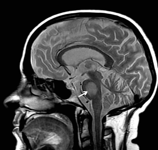
Osmotic demyelination syndrome (ODS)
In chronic hyponatremia, brain cells extrude organic osmolytes to avoid swelling. Those take days to replace, so if the sodium rises quickly the adapted cell is left hypertonic to its surroundings, shrinks, and the myelin-maintaining oligodendrocytes are injured. It is a complication of the treatment, not of the hyponatremia.
- Where. Classically the base of the pons — the old name central pontine myelinolysis — but also basal ganglia, thalamus and cerebellum. A normal pons does not rule it out.
- When. Biphasic: the patient improves as the sodium normalizes, then deteriorates 1 to 14 days later. MRI can lag by three weeks, so a normal early scan does not exclude it.
- How it presents. Dysarthria and dysphagia, then confusion, quadriparesis, pseudobulbar palsy, and at worst a locked-in state. Extrapontine disease can look like delirium.
- Who is at risk. Sodium below 105 mmol/L, hypokalemia, alcohol use disorder, malnutrition, advanced liver disease.
- What happens to them. Across 541 reported patients, 51.9% recovered well and 24.8% died — so a severe presentation is not by itself a reason to withdraw care. No specific treatment exists; prevention is the whole point of the limit.
Two things raise the sodium without anyone ordering it, and both count toward the daily ceiling. Replacing potassium raises the sodium, because potassium entering cells displaces sodium outward — a common and invisible contributor in exactly the malnourished, hypokalemic patients most at risk of demyelination. And correcting hypovolemia switches ADH off, after which the kidney dumps free water on its own. If the urine output suddenly climbs and the urine looks dilute, the sodium is about to rise faster than your infusion explains.
If you overcorrect, it is reversible — act on it. Stop the hypertonic saline, give 5% dextrose to replace free water, and add desmopressin 2–4 µg IV to stop the ongoing loss. Escalate, but recognizing it at 03:00 and stopping the offending fluid is a clerk-level action.
The limit is genuinely contested, and you should know why. Three large observational studies — MacMillan (22,858 admissions across five Toronto hospitals), Seethapathy (3,274 patients) and Kinoshita (1,024 ICU patients) — all found ODS rare and slower correction associated with higher mortality. In the Toronto cohort ODS occurred in 12 patients (0.05%), and 7 of those 12 had not been corrected rapidly. But sicker patients are corrected more slowly because they are sicker, which is the confounding these designs cannot remove, and 64% of that cohort was never imaged. The NephMadness 2024 hyponatremia region argues both sides at length.
What this changes for you: nothing yet, and that is the point. No guideline has moved, so the 8–10 mmol/L limit is what you follow. What it should change is the reflex — most ODS in these cohorts happened to patients who were not corrected quickly, which suggests the host matters at least as much as the rate.
5. On the Ward
Estimated read time: 3 minutes
Put the monitoring interval in the order, not just the fluid. A sodium being corrected needs q2–4h checks at first, stretching to q6h once the trajectory is stable. Hypertonic saline ordered without a rechecking schedule is how overcorrection happens.
Hand over the ceiling, not just the number. Correction crosses shift changes, so say all three parts out loud: “Sodium 118, up 6 from the nadir, ceiling 122 by 22:00 tonight.” A number on its own leaves the next person to reconstruct the arithmetic.
Count the diluent. Restriction prescribed but not enforced — the patient drinking from the tap, antibiotics arriving in 250 mL bags — is a common reason SIADH appears not to respond.
Review the medication list at discharge. If a thiazide or an SSRI caused this, restarting it undoes the admission.
Hypernatremia, briefly — the mirror image
A sodium above 145 mmol/L is almost always a water deficit in someone who cannot get to water themselves: intubated, living with dementia, post-operative with no oral intake. The sodium marks that access problem as much as a disorder in its own right.
- Estimate the deficit: total body water × (serum sodium ÷ 140 − 1), total body water being roughly 0.6 × weight in kg for men and 0.5 × for women
- Replace it enterally where possible — water by mouth or nasogastric tube beats intravenous D5W
- The same 10 mmol/L per 24 hours limit applies in reverse — dropping the sodium too fast causes cerebral edema
- Check for diabetes insipidus only if the urine stays dilute despite a rising serum sodium
Related Podcasts
Test Yourself
Click on your answer to reveal the explanation.
A 74-year-old woman is on day 3 of an admission for community-acquired pneumonia. She is comfortable, her blood pressure is 128/74 mmHg and has been stable, and she is not orthostatic. She has no edema and a normal jugular venous pressure. Morning bloodwork shows a sodium of 126 mmol/L, down from 138 on admission. Serum osmolality is 266 mOsm/kg, urine osmolality 505 mOsm/kg and urine sodium 62 mmol/L. Glucose, TSH and a morning cortisol are normal. She takes sertraline. She has no headache, nausea or confusion.
What is the most appropriate initial management?
A 52-year-old man with alcohol use disorder is admitted with a serum sodium of 106 mmol/L and a witnessed seizure. He receives two 150 mL boluses of 3% saline overnight, the seizures stop, and he is now alert and oriented. His blood pressure is 122/70 mmHg and stable. Twenty hours after presentation his sodium is 119 mmol/L. Urine output over the past four hours has been 900 mL of dilute urine. His potassium was 2.9 mmol/L on admission and has been replaced.
What is the most appropriate next step?
Reflect
The three studies in section 4 all point the same way: osmotic demyelination is rarer than taught, and slower correction was associated with worse outcomes, not better. No guideline has changed, and you are still expected to cap correction at 8–10 mmol/L in 24 hours. Describe how you would explain that tension to a patient’s family — that you are deliberately correcting slowly, using a limit that recent evidence has called into question.
Please do not include patient identifying details — no names, initials, dates, medical record numbers, or any detail that could identify a specific patient or encounter. Write about your reasoning, not about a real person.
Further Reading
- GuidelineClinical practice guideline on diagnosis and treatment of hyponatraemia (ERBP/ESICM/ESE, 2014)
- GuidelineDiagnosis, evaluation, and treatment of hyponatremia: expert panel recommendations (2013)
- Cohort studyOsmotic Demyelination Syndrome in Patients Hospitalized with Hyponatremia (NEJM Evidence, 2023)
- Randomized trialRapid intermittent bolus vs slow continuous infusion of hypertonic saline — the SALSA trial (2021)
- BlogNephMadness 2024: Hyponatremia Region — both sides of the correction-rate argument
Week 3